David K. Joyce, Newmarket, Ontario
Dr. Donald V. Doell, Grafton, Ontario
Introduction
In 2016, David K Joyce (DKJ) met up with co-author Dr. Donald V. Doell (DVD) at the annual convention of the Prospectors and Developers Association of Canada (PDAC). The PDAC is a gathering of approximately 28,000 people, mostly geologists, engineers, mining and exploration company executives, investors, and other interested and related people. There are many activities at the PDAC, one of the most interesting being the “Investors’ Exchange”, where people can visit booths of hundreds of mining and exploration companies to learn technical details of their deposits, mineral exploration strategies and/or investment potential.
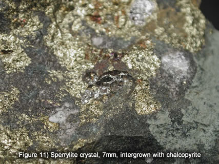
DVD and DKJ were both there visiting various companies looking at investment potential and, of course, potential for mineral specimen acquisition! There are usually rock/ore or mineral specimens at each booth representing the mineralogy and geology of various deposits. One booth, in particular, was of interest to us. Wallbridge Mining Company (Wallbridge) had operated a small mine north east of Sudbury for platinum-group elements, copper and nickel. During the exploration and subsequent mining stages, Wallbridge had recovered excellent specimens of sperrylite crystals in matrix. Usually, the sperrylite crystals were embedded in chalcopyrite/pyrrhotite matrix but sometimes they were embedded in quartz, associated with epidote. Please see another article on this website, https://djoyceminerals.com/pagefiles/articles_brokenhammermine.asp that gives more details about the Broken Hammer Mine, its geology and minerals.
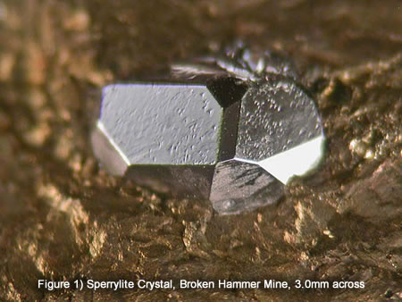
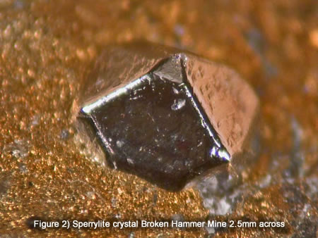
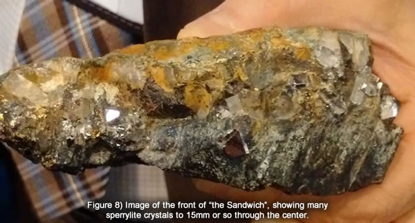
Wallbridge Mining Co. had one particular specimen that had been nicknamed “the Sandwich”, since it appeared to be a rich specimen of chalcopyrite with a layer of large sperrylite crystals in the center( the meat) between outer layers of golden chalcopyrite and rock (the bread). Mine management speculated whether, or not, the sperrylite crystals were not only observable at the front edge of the specimen as a line or were they a discontinuous plane inwards between the layers of chalcopyrite? How to tell without splitting or breaking “the Sandwich”?
DVD thought that it could be possible to “see inside” the specimen, utilizing the advanced x-ray imaging offered by CT Scanners, the type used in hospitals. Of course, CT scanners are usually used for imaging relatively low density internal organs of medical patients. However, we wondered if it would be possible to modify soft-tissue x-ray techniques with a CT Scanner to examine the insides of a much higher density material, a piece of chalcopyrite (density of 4.2 g/cc) that contained sperrylite crystals (density of 10.55 g/cc). It was an interesting discussion and excited our imaginations.
Sperrylite Specimens
A couple of months later, A. Soever, Chairman of Wallbridge Mining, contacted DKJ and asked him if he would prepare and sell some sperrylite crystal specimens on behalf of Wallbridge Mining Co. Subsequently, DKJ took possession of a number of pieces of sperrylite-bearing chalcopyrite. A couple of them were large and had only broken sperrylite crystals exposed on the outside. It occurred to DKJ that it might be a good idea to examine these bigger chunks, internally, using a CT Scanner, as suggested by DVD. He contacted DVD and discussed the matter. DVD, has very good relations with the Montreal Neurological Institute (MNI), at Montreal, Quebec, so he contacted colleagues there and discussed with them, whether such x-ray technique could work on such specimens and was the MNI willing to allow us to try this technique on the Wallbridge specimens. They were agreeable! We contacted Wallbridge and they also agreed to allow us to image ”the Sandwich” to see if sperrylite crystals existed inside the chalcopyrite, in addition to what was observable on the outside.
CT Scanner
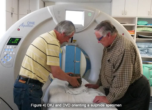
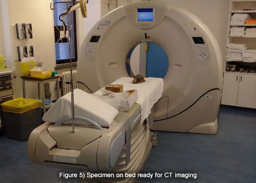
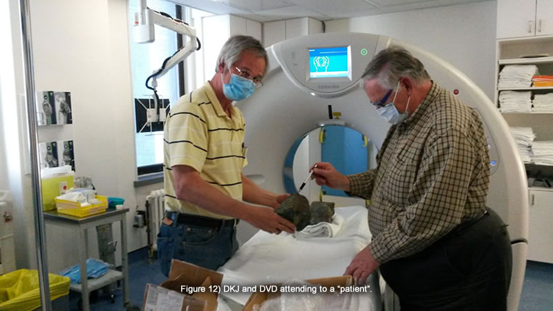
The MNI agreed to allow us to use their scanner, a Toshiba Aquilon ONE CT scanner, for a short period of time, when it was not required for medical examinations.
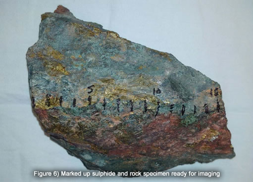
DKJ marked the larger specimens with 1.0cm increments in an attempt to better understand where the sperrylite crystals were later during trimming operations. The idea was to try to orient the fragments in concert with the x-ray images afterwards in an effort to locate any sperrylite crystals as accurately as possible for successful trimming and exposure.
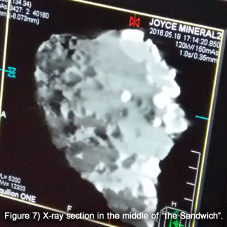
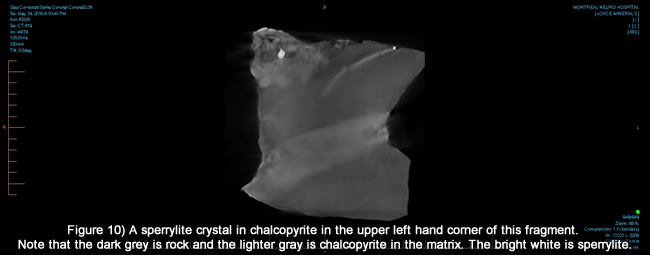
The MNI Technician was made available to us and very quickly adjusted voltages and other parameters to internally image chunks of chalcopyrite. It was agreed to image the pieces in 1mm “slices”, that is, an image of the internal structure of the specimens was captured perpendicular to x,y and z axes every one mm. This allowed us to look inside the specimens, in the same way that you might flip the pages of a book with 1mm thick pages –but in three dimensions! Instantly, we could see that most of the specimens were barren of sperrylite crystals except that every once in a while, a bright white area would appear in an otherwise grey mass - a sperrylite crystal!
As it turned out, the sandwich did NOT have many more sperrylite crystals extending into it in a plane. It looks like a sandwich but, in fact, it is not. There are a couple of buried sperrylite crystals, though, as you can see in the x-ray image of the specimen. DKJ (sadly) returned the Sandwich to Wallbridge Mining Company where you can see it at their office or at select trade shows. It is quite something! If Wallbridge ever DID decide to break it up to better expose the various sperrylite crystals, we totally understand the inner structure and location of every crystal. It would be great challenge to prepare several specimens from it!
Specimen Preparation
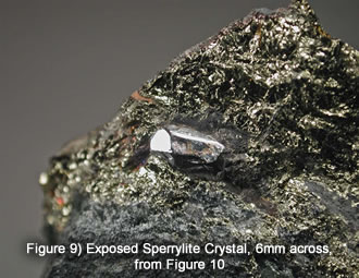
Unfortunately, the sperrylite crystals were mostly the kind that are intergrown with the chalcopyrite and rock. As such, most just split irregularly, revealing crystal sections, impregnated with chalcopyrite, rather than separating cleanly as solid crystals do from massive sulphides. One did turn out fairly well. It was a cube modified by octahedral and pyritohedral(?)crystal faces. Despite knowing where the crystals are in the matrix, we are still subject to the vagaries of how the rock and massive will actually fracture. It rarely actually breaks exactly how you expect or want!
So, even though the CT imaging examination worked and we had a good idea where the sperrylite crystals were in these specimens, they were not that many and those that were there were not all that amenable to preparation and exposure. If they were the kind that are solid, discrete from and separated from the chalcopyrite easily, our experiment may have been a more commercially successful.
Acknowledgements and Thank You
We would like to thank the Walbridge Mining Co., particularly Alar Soever, Chairiman and Marz Kord, President, on a couple of counts, particularly for enabling the preservation of sperrylite crystal specimens from the Broken Hammer Mine and for being encouraging about the concept of CT scanner analyses and for allowing us to borrow “the sandwich” for these purposes.
We sincerely appreciate the generosity and cooperation of Dr. D Tampieri and A. Hatem, both of the Montreal Neurological Institute and Hospital, at Montreal.
Thank you to Violet Doell for the great photo’s that she took of us working at the Montreal Neurological Institute.